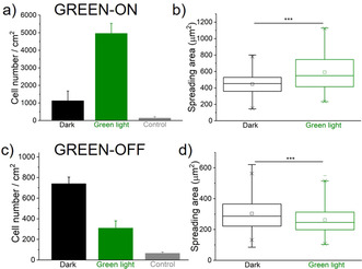
Figure 2.

Cell adhesion on GREEN‐ON (a, b) and GREEN‐OFF (c, d) surfaces. Quantification of a), c) the number of cells that adhere based on the nuclear DAPI staining and b), d) spreading area of the cells based on the actin staining. The surfaces were either kept in the dark or illuminated with green light for 5 minutes prior to seeding cells. A surface without protein functionalization was used as a control. One Way ANOVA test, p‐value ***<0.001.
