
Figure 1.
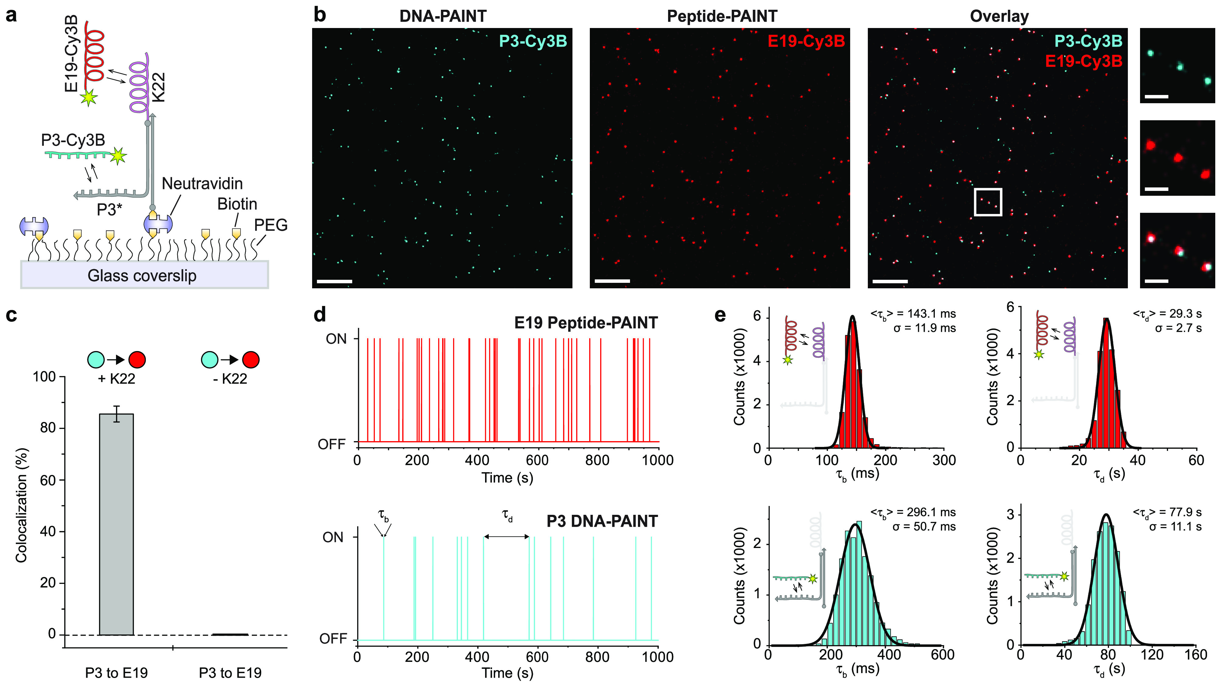
Characterization of transient coiled coil interactions using single-molecule imaging. (a) Schematic diagram depicting the single-molecule imaging assay for characterizing Peptide-PAINT. A peptide coil (K22) is conjugated to a DNA strand (DBCO-S1HP3H, see Table S4) for hybridization to a surface-bound complement. The strand on the coil is furthermore extended with a DNA-PAINT docking sequence for direct visualization. (b) Representative single-molecule localization data resulting from DNA-PAINT (left, cyan), Peptide-PAINT (middle, red), and their overlay (right) of a selected field of view. Zoom-ins highlight colocalization of DNA- and Peptide-PAINT. (c) Colocalization analysis (from DNA- to Peptide-PAINT signals) yields 86 ± 3% (mean and standard deviation) for the positive control and no colocalization for the negative control, which lacks the K22 peptide (n > 1500). (d) Exemplary binding traces showing transient interactions of E19 with surface immobilized K22 and P3 binding to its corresponding docking strand (with highlighted bright and dark times τb and τd). (e) τb and τd distributions for E19 (n = 18938) and P3 (n = 15544) and corresponding Gaussian fits (mean and standard deviation are stated). Scale bars: 300 nm (b, overview), 50 nm (b, zoom-ins).