
Figure 2.
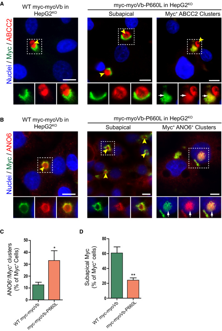
The MVID‐associated myoVb‐P660L mutation causes the intracellular accumulation of canalicular proteins. (A,B) Immunofluorescent microscopy images of myc‐tagged myoVb proteins, ABCC2 and ANO6, in HepG2KO expressing myc‐myoVb and myc‐myoVb‐P660L. HepG2KO cells expressing myoVb‐P660L show intracellular colocalization of myc and ABCC2 (white arrows). Yellow arrowheads indicate BCs. Scale bars: 10 μm. (C) Quantification of the percentage of myc‐positive cells that show intracellular clusters/accumulations of myc localized with ANO6. (D) Quantification of the percentage of myc‐positive cells that show subapical localization of myc‐tagged myoVb proteins.