Figure 7.

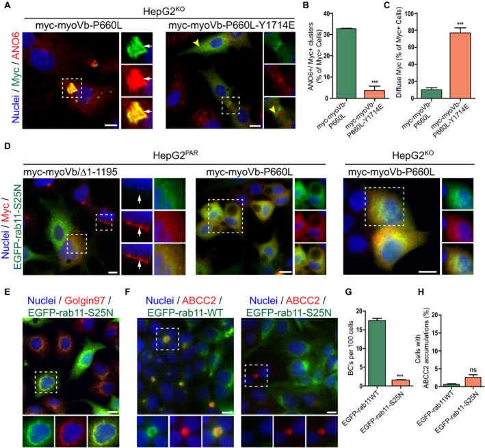
The disrupting effect of motorless myoVb on canalicular protein localization requires active rab11a. (A) ANO6 and myc labeling in HepG2KO expressing myc‐myoVb‐P660L or myc‐myoVb‐P660L‐Y1714E. Myc‐myoVb‐P660L frequently accumulated intracellularly with ANO6 (white arrows), whereas myc‐myoVb‐P660L‐Y1714E appeared diffuse in the cytoplasm or subapical (yellow arrowheads). (B) Quantification of the percentage of myc‐positive cells that show intracellular clusters/accumulations of myc localized with ANO6 in HepG2KO cells expressing myc‐myoVb‐P660L or myc‐myoVb‐P660L‐Y1714E. (C) Quantification of the percentage of myc‐positive cells that show subapical localization of myc in HepG2KO cells expressing myc‐myoVb‐P660L or myc‐myoVb‐P660L/Y1714E. (D) HepG2 coexpressing EGFP‐rab11aS25N and myc‐myoVb/Δ1‐1195 in HepG2Par cells (left), EGFP‐rab11aS25N and myc‐myoVb‐P660L in HepG2Par cells (middle), and EGFP‐rab11aS25N and myc‐myoVb‐P660L in HepG2KO HepG2 (right). In all three conditions, the presence of EGFP‐rab11aS25N prevented the clustering of the respective myoVb mutants. (E) EGFP‐rab11aS25N colocalized with golgin‐97 in HepG2 cells. (F) ABCC2 labeling in HepG2 overexpressing wild‐type EGFP‐rab11a or EGFP‐rab11aS25N. In EGFP‐rab11aS25N transduced cells, BC formation was only seen in cells with no or very low expression of the construct (right side, box). (G) Quantification of BC formation (expressed as BC/100 cells) in HepG2 cells expressing wild‐type EGFP‐rab11a or EGFP‐rab11aS25N. (H) Quantification of the percentage of HepG2 cells showing accumulation of ABCC2 on expression of wild‐type EGFP‐rab11a or EGFP‐rab11aS25N.
