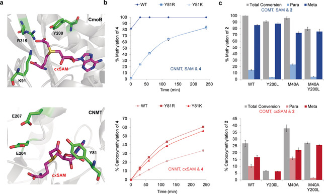
Figure 3.

a) Crystal structure of CmoB and a model of cxSAM binding into the CNMT active site. Top: The CmoB active site highlights three residues thought to interact with the carboxymethyl group (PDB ID: 4QNU). Bottom: WT CNMT with cxSAM positioned in the active site (based on PDB ID: 6GKV, which has SAH bound), highlighting three residues predicted to be in proximity to the carboxymethyl group. b) CNMT time courses with THIQ 4 (0.25 mm) and SAM or cxSAM were conducted using either 1 mm SAM (top/blue) or 1.6 mm cxSAM (bottom/red), with commercial SAM comprising only the active S enantiomer at 80 % purity and cxSAM being synthesised as a mixture of enantiomers. c) COMT activity assays with catechol 2 (0.2 mm) and SAM or cxSAM were conducted for 5 h at 37 °C to compare % methylation and carboxymethylation, using either 1 mm SAM (top/blue) or 1.6 mm cxSAM (bottom/red). Assays were done in triplicate and standard errors calculated.
