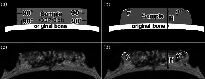
FIGURE 1.

Radiomorphometric measurement of the height and the angles of the upper margin of the implanted octacalcium phosphate and collagen composite (OCP/Col). The height (H) between the bottom surface of the original bone and the angles of upper margin (θ and θ′) of the implanted OCP/Col was measured 4 and 12 weeks after implantation by using micro‐computed tomography (micro‐CT) image. To measure the angles, the apex of the implant at 4 or 12 weeks was plotted on the micro‐CT image, and two inflection points on both side of the apex were plotted on a ridgeline. After that the angle centered around the apex was measured. (a) Schema of immediately after implantation. (b) Schema of 12 weeks after implantation. (c) Micro‐CT image of 12 weeks after implantation. (d) Micro‐CT image with description of the height and upper angles
