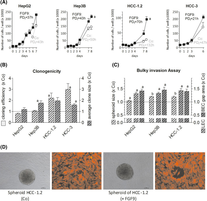
FIGURE 1.

FGF9 enhances growth, cloning efficiency and the invasive phenotype of hepatoma/hepatocarcinoma cells. (A), Twenty‐four h after seeding cells were treated with 10 ng of FGF9/ml medium. Cells were counted at regular intervals; the population doubling time (PDt) was calculated using the first and last count. (B), Cell lines were plated at low densities and were treated with 10 ng of FGF9/ml medium. Clone numbers and size were determined after 10 d (HepG2) or 2 wk (Hep3B, HCC‐1.2, HCC‐3). (C,D), Spheroids were formed (for details see Methods) and were placed on confluent LEC and BEC monolayers for~4 h. Pictures of spheroids and gaps in monolayers underneath were taken and sizes were measured by ImageJ software. (D), Representative spheroids (in gray) of HCC‐1.2 cells and corresponding gaps formed beneath in LEC (red fluorescence). (A‐C), All data are given as means ± SEM of ≥3 independent experiments. Statistics in (A) by paired t‐test for FGF9 vs untreated control (Co) on the last day: a, P < .05; statistics in (B) and (C) by One Sample t‐test for FGF9 treatment vs Co: a, P < .05; b, P < .01
