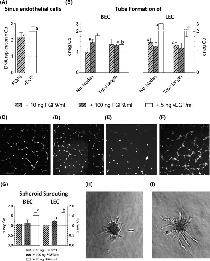
FIGURE 2.

FGF9 stimulates neoangiogenesis. (A), Freshly isolated rat liver sinus endothelial cells were kept under standard conditions, as described, 14 and were treated with 10 ng of FGF9/ml or 5 ng vEGF/ml medium; 24 h later DNA replication was determined by 3H‐thymidine incorporation and scintillation counting. (B‐F), Tube formation of BEC and LEC was induced by treatment with FGF9 for 14 h, as described in methods. BEC: untreated (C) or treated with 10ng FGF9/ml (D); LEC: untreated (E) or treated with 100ng FGF9/ml (F). (G‐I), Spheroid sprouting assay was performed as described in methods. Sprouting BEC spheroid, being untreated (H) or treated with 100 ng of FGF9/ml medium (I). Data in (A), (B) and (G) give means ± SEM of ≥3 three independent experiments. Statistics by One Sample t‐test: a, P < .05; b, P < .01
