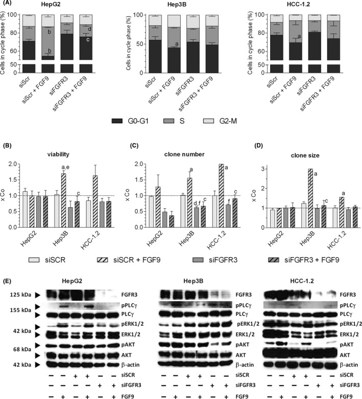
FIGURE 5.

siFGFR3 antagonizes the FGF9‐mediated effects on cell cycle, viability, clone formation and signalling. (A‐D), Cells were transfected with siSCR or siFGFR3 (4392421/s5167, ThermoFisher Scientific) and re‐seeded in medium containing 1% FCS 24 h after transfection. Four hours later cells were treated with 10 ng FGF9/ml medium. Further methodical details see Table S6. (A), FACS determined the relative cell cycle distribution 48 h after FGF9 treatment. (B), Number of viable cells was determined by the MTT assay. (C‐D), Clones were fixed and stained with crystal violet after ~ 14 d. Number and size of clones were quantified by ‘LUCIA G image analysis software’. (E), 20 min after FGF9 stimulation, protein was isolated, separated on 10% SDS‐gels, and immunoblotted. (A‐D), Data are expressed as mean ± SEM of 3 independent experiments. Statistics by unpaired t‐test: any treatment without FGF9 vs any treatment with FGF9: a, P < .05; b, P < .01; any treatment without siFGFR3 vs any treatment with siFGFR3: c, P < .05; d, P < .01. Statistics by One Sample t‐test for any treatment vs control: e, P < .05; f, P < .01
