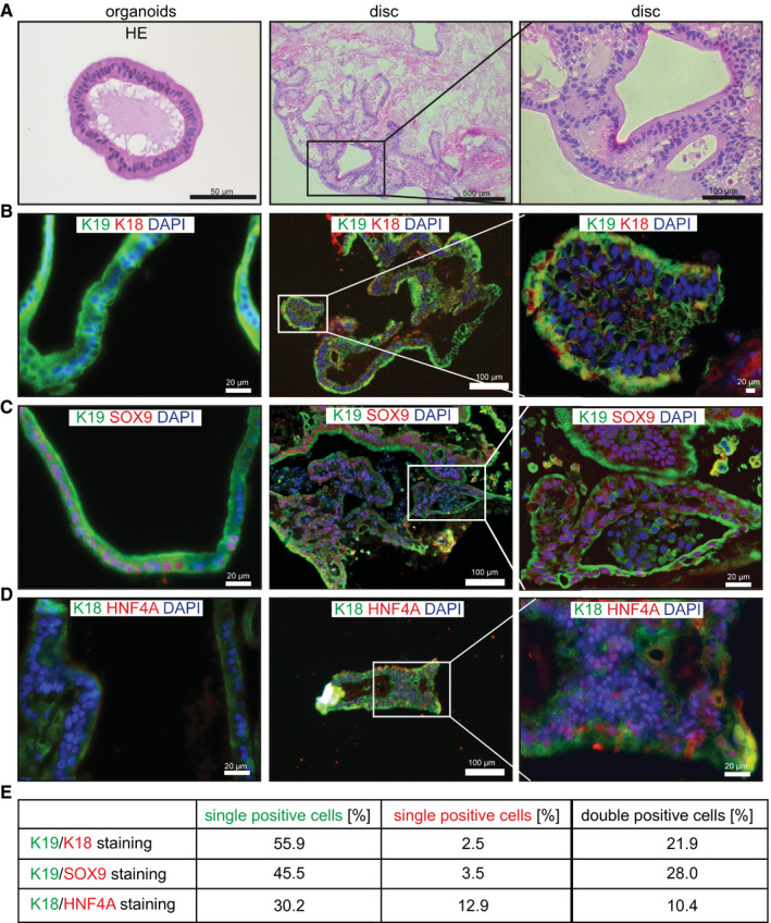
Figure 3.

Tissue formation of spinner flask organoids on liver ECM. (A‐D) Organoids were expanded in spinner flasks for 14 days and then seeded on decellularized rat liver discs. Reseeded discs were cultured for 2 days in EM supplemented with BMP‐7 and then differentiated for 5 days in human organoid DM. Four different donors (2‐3 discs per donor) were analyzed in independent experiments. HE stainings (A) and immunofluorescent analysis (B‐D) of paraffin‐embedded organoids and liver discs are shown. Note that K18+ and HNF4a+ cells were not present in the organoids at the time of seeding on decellularized discs. (E) Quantification of the different cell populations. Abbreviation: HNF4a+, hepatocyte nuclear factor‐4‐alpha.
