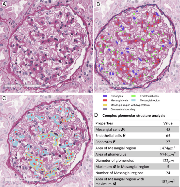
Figure 3.

Assessment of internal glomerular structures. (A) Original glomerulus image with PAS stain. (B) Intrinsic glomerular cell segmentation, with detected mesangial cells (M), podocytes (P), endothelial cells (E), and glomerulus boundary. (C) Assessment of mesangial regions, including mesangial regions and mesangial cells. (D) Measurement results of the glomerulus in C.
