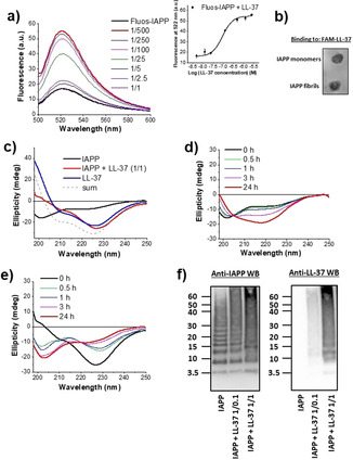
Figure 2.

Characterization of the LL‐37‐IAPP interaction. a) Determination of the app. K d by fluorescence spectroscopic titrations. Fluorescence emission spectra of Fluos‐IAPP (5 nm) alone or with various amounts of LL‐37 (pH 7.4) as indicated. Inset: binding curve (mean±SD, 3 titration assays). b) Binding of FAM‐LL‐37 to IAPP monomers and fibrils as determined by DB. IAPP monomers and fibrils (40 μg) were spotted on a nitrocellulose membrane and probed with FAM‐LL‐37 (200 nm; results representative of 4 assays). c) Far‐UV CD spectra of IAPP (5 μm), IAPP‐LL‐37 (1:1; 5 μm each), and LL‐37 (5 μm, 0 h,pH 7.4). The sum of the spectra of LL‐37 and IAPP is also shown. d, e) Kinetic follow‐up of IAPP misfolding alone (d) or its 1:1 mixture with LL‐37 (e) through far‐UV CD spectroscopy. Conditions as in (c). f) Characterization of IAPP/LL‐37 hetero‐assemblies through cross‐linking with glutaraldehyde (pH 7.4), NuPAGE, and western blotting (IAPP 30 μm; IAPP/LL‐37 1:0.1 or 1:1). A representative gel (n>5) is shown.
