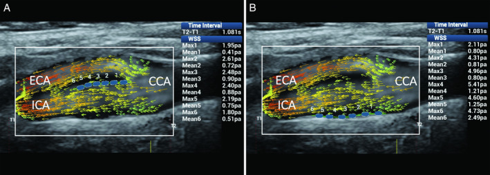
Figure 8.

Mainly laminar flow in the CB. Ultrasound vector flow imaging shows the vector direction remaining parallel to the vessel walls. Wall shear stress measurements (blue dots) gave maximum and mean values within the normal range: in particular, along the flow divider, 1.80 to 2.61 and 0.41 to 0.90 Pa, respectively (A), and along the opposite wall from the flow divider, 2.11 to 5.41 and 0.80 to 2.49 Pa (B). CCA indicates common carotid artery; ECA, external carotid artery; and ICA, internal carotid artery.
