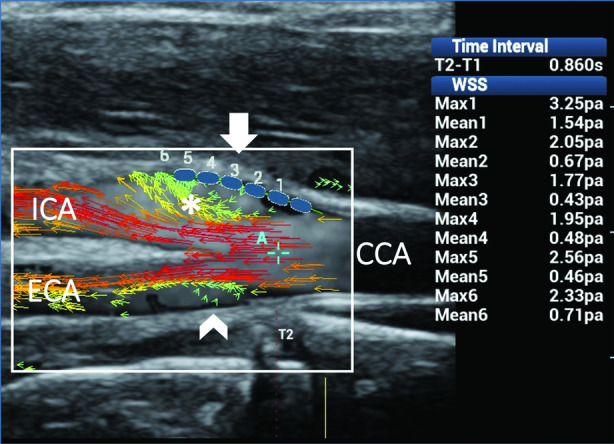
Figure 9.

Rotational flow in the internal carotid artery (ICA). Ultrasound vector flow imaging shows flow layer separation in both the ICA sinus and external carotid artery (ECA). High‐velocity red vectors move along the 2 sides of the flow divider. Low‐velocity short green vectors rotate around an axis of flow along the outer wall while moving forward in the ICA (asterisk) and move back in a small recirculation area along the outer wall in the ECA (arrowhead). Wall shear stress measurements (blue dots) along the outer wall of the ICA (arrow) gave maximum and mean values within the normal range (1.77–3.25 and 0.43–1.54 Pa, respectively), thus confirming the steady shear stress related to the helical flow. CCA indicates common carotid artery.
