Figure 2.

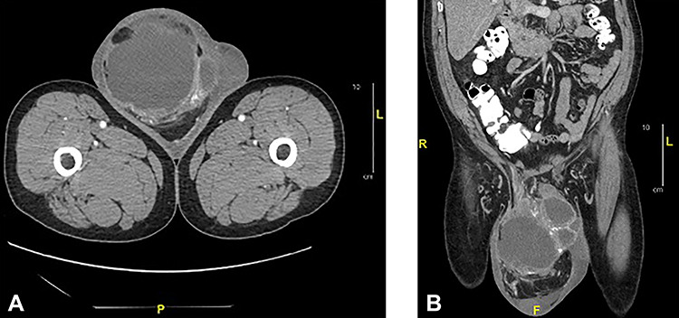
Axial (a) and sagittal (b) CT of abdomen and pelvis with oral and intravenous contrast: showed a large complex cystic and solid mass measuring 12.0 x 15.5 x 19.0 cm in the right scrotum.

Axial (a) and sagittal (b) CT of abdomen and pelvis with oral and intravenous contrast: showed a large complex cystic and solid mass measuring 12.0 x 15.5 x 19.0 cm in the right scrotum.