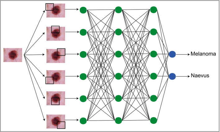
Figure 3.

Schematic depicting how classification tasks are performed in convolutional neural networks. Pixel data from an image are passed through an architecture consisting of multiple layers of connecting nodes. In convolutional neural networks, these layers contain unique ‘convolutional layers’, which operate as filters. These filters work because it was recognized that the location of a feature within an image is often less important than whether that feature is present or absent – an example might be (theoretically) the presence or absence of blue‐grey veiling within a melanoma. A convolutional ‘filter’ learns a particular feature of the image irrespective of where it occurs within the image (represented by the black squares). The network is composed of a large number of hierarchical filters that learn increasingly high‐level representations of the image. These could in principle learn dermoscopic features similar to those described by clinicians, although in practice the precise features recognized are likely to differ from classic diagnostic criteria.
