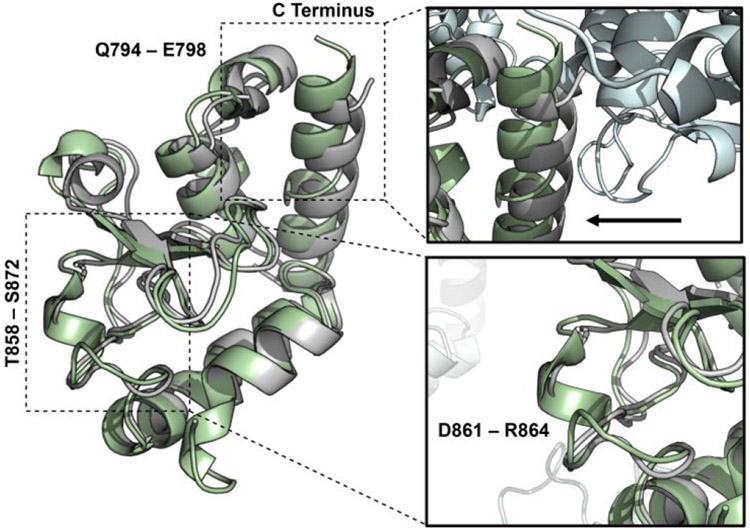
Figure 2. X-ray structure of HNH.
The X-ray structure of the isolated HNH domain (PDB code: 6O56, green), solved at 1.30 Å resolution, is overlaid with the X-ray structure of the HNH domain from the full-length S. pyogenes Cas9 (PDB code: 4UN3, gray).5

