Figure 5.

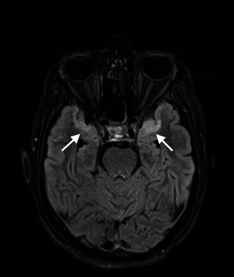
MRI of the brain T2-fluid attenuated inversion recovery axial image revealing hyperintense, rather symmetrical, signals in the anteromedial temporal lobes bilaterally.

MRI of the brain T2-fluid attenuated inversion recovery axial image revealing hyperintense, rather symmetrical, signals in the anteromedial temporal lobes bilaterally.