
Figure 3.
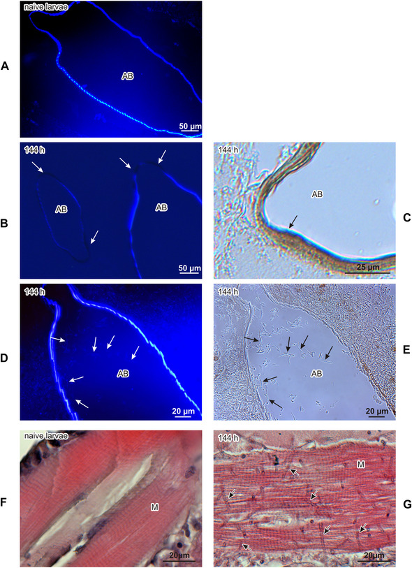
Air bags of healthy (A) and infected (B–E) G. mellonella larvae. (A, B, D) Staining with calcofluor‐white, observed under UV light; (C, E) no staining, seen under visible light. (A) Air bag of a healthy larva. (B) Disturbed walls of air bags have no ability to bind calcofluor‐white (indicated by arrows). (C) Melanized wall of air bag is indicated. (D) Calcofluor‐white binds to chitin lining the walls of the air bags and to chitin present in the thin cell walls of B. bassiana (indicated by arrows) growing inside the air bags. (E) Hyphae of B. bassiana (indicated by arrows) seen under visible light. Muscles of healthy (F) and infected (G) G. mellonella larvae (144 h after natural infection). A growing fungus is indicated by arrows. AB, air bags; M, muscles.