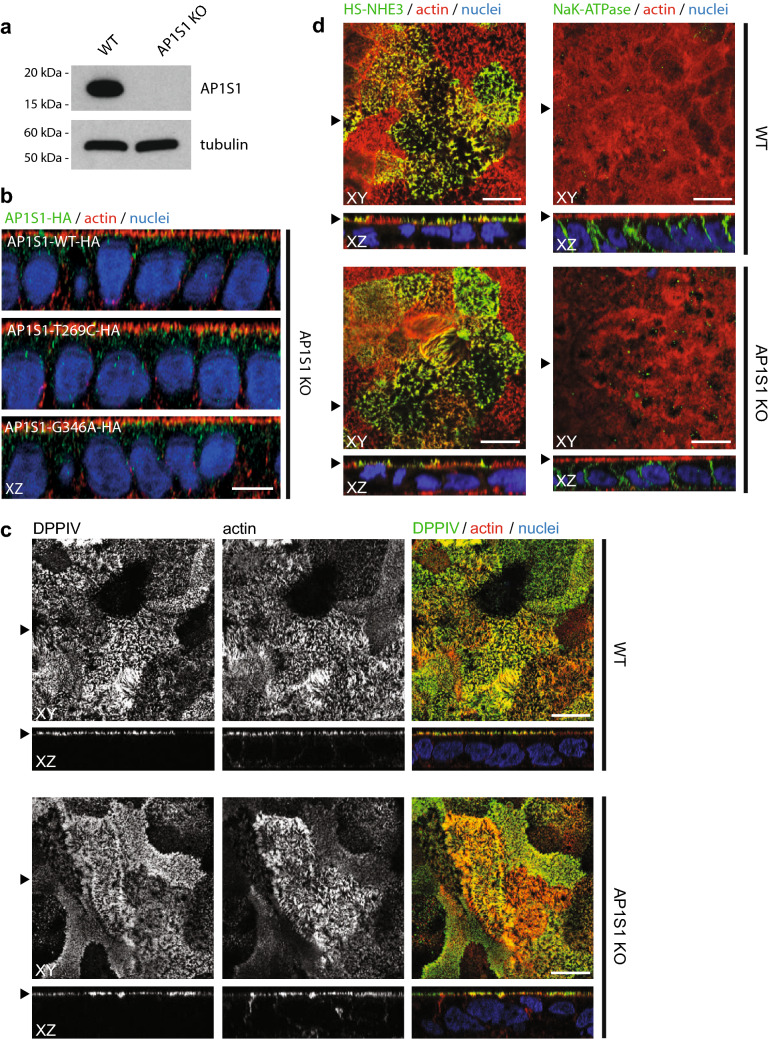
Fig. 2.
a Western blot detects AP1S1 in CaCo2 WT but not in CaCo2-AP1S1-KO cells. Tubulin is used as loading control. b Laser-scanning immunofluorescence micrographs (LSM) of CaCo2-AP1S1-KO cells expressing AP1S1-WT-HA, AP1S1T269C-HA or AP1S1-G346A-HA. Vesicular localization pattern of AP1S1. Scale bar 10 µm. c LSM of CaCo2 WT and AP1S1 KO cells. DPPIV staining, microvilli morphology (actin) and NHE3 apical localization (d) is unaffected by AP1S1 depletion. Basolateral localization of NaK-ATPase is not affected by loss of AP1S1. Scale bar 10 µm, arrowheads indicate respective plane in XY and XZ

