Fig. 5.
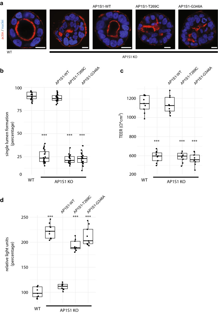
a Epithelial cysts formed by CaCo2 cells grown in Matrigel. Loss of AP1S1 or expression of mutated AP1S1 results in no or multiple lumina (actin staining) formation indicative of disturbed epithelial polarity. Scale bar 10 µm. b AP1S1 genotype-dependent central lumen formation (dot box plot, Mann–Whitney U test ***p < 0.001, three independent biological experiments per condition, a total of 150 cysts per condition were assessed). c Loss of AP1S1 without or with expression of patient mutations results in a significantly decreased transepithelial electrical resistance (TEER) in CaCo2 monolayers as compared with AP1S1 wild-type re-expression in CaCo2-AP1S1-KO cells (dot box plot, Mann–Whitney U test ***p < 0.001, three independent biological experiments per condition). d Epithelial barrier function for Dextran-TexasRed passage was tested by measuring fluorescence after ten hours in lower medium chamber. Increased fluorescence, e.g., increased monolayer permeability, was observed upon loss of AP1S1 or expression of patient mutations. Relative light units were normalized to WT measurements (dot box plot, Mann–Whitney U test ***p < 0.001, three independent biological experiments per condition)

