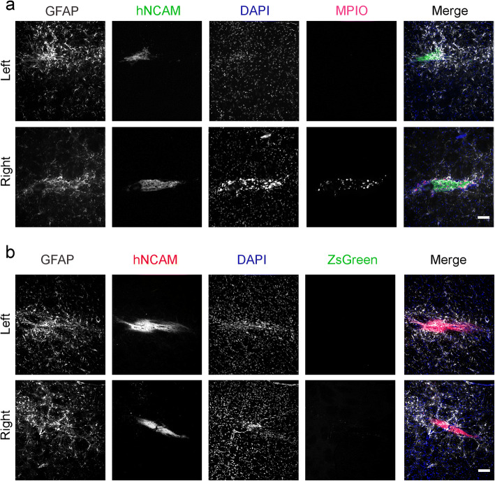
Fig. 5.
Glial reaction at the injection sites. a Immunofluorescence microscopy of brains from rats that received MPIO-labelled DAPCs (left hemisphere: unlabelled; right hemisphere: labelled). The presence of human cells is identified with hNCAM staining, and the intensity of GFAP staining is stronger in these areas. MPIOs are only seen in the right hemisphere. b Immunofluorescence microscopy of brains from rats that received Fluc-ZsGreen+ DAPCs (left hemisphere: untransduced control cells; right hemisphere Fluc-ZsGreen+ cells). Scale bars correspond to 100 μm.

