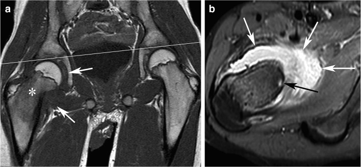
Fig. 1.
Painless OO in a 13-year-old boy with a painless limp and normal spinal MRI (not shown) requested for the limping gait. A coronal T1-weighted image of the pelvis (a) shows abduction of the right femur relative to the pelvic plane (line), low signal intensity in the bone marrow of the right femoral neck (asterisk), and synovial swelling with low signal intensity (arrows). An axial fat-saturated proton density-weighted MR image (b) shows both synovial swelling with high signal intensity (white arrows) and the OO nidus in the femoral neck (black arrow), which was confirmed by a CT scan (not shown). The OO was then successfully treated by radiofrequency ablation

