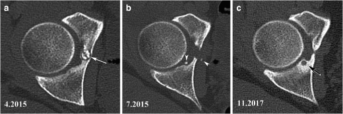
Fig. 10.
Recurrence near the original site of a resected OO. An OO involving the deeper aspect of the acetabulum (arrow in CT image in a) was arthroscopically resected. A CT examination performed after surgery (b) showed complete lesion resection and the presence of small bone fragments (arrowheads) near the resected site. The pain recurred after several months and repeated CT examination performed 30 months after surgery demonstrated appearance of a small lucent area surrounded by hyperostosis near the resection site (arrow in c). Recurring OO nidus was confirmed on DCE-MRI (not shown) and successfully electrocoagulated

