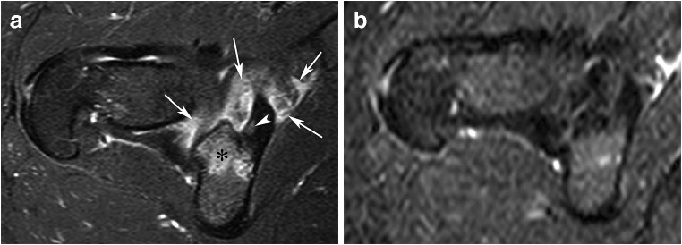
Fig. 12.
Resolution of an OO (not proved histologically) of the inferior margin of the acetabulum in a 24-year-old woman with nocturnal hip pain from several months. A bone scintigraphy showed increased focal absorption in the lower part of the acetabulum (not shown). Axial fat-saturated T2-weighted MR image (a) shows edema-like bone marrow in area behind the acetabulum (asterisk) and significant soft tissue edema (arrows). A small oval bone defect with intermediate signal (arrowhead) is present within a sclerotic zone of the acetabulum and corresponds presumably to an OO nidus. Because an excellent response to NSAIDs, medical treatment was proposed. Two years later, the pain had significantly decreased and an MRI showed a marked regression of the edema around the unchanged nidus (not shown). Six years later, the patient was totally asymptomatic and MRI (b) demonstrated complete abnormalities resolution without surgery

