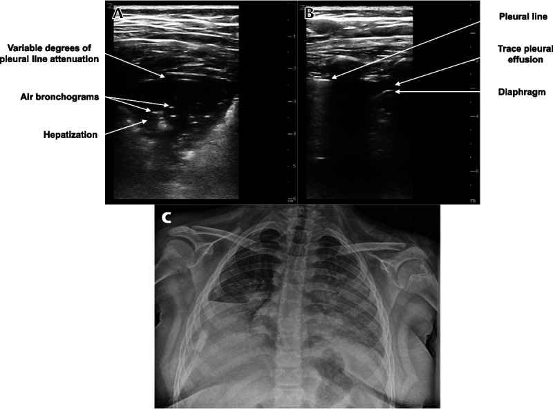
FIGURE 1.

Lung POCUS and chest radiograph of a 9-year-old girl with respiratory distress. A, Transverse image of a right posterior lung field showing a consolidation with air bronchograms and variable degrees of pleural line attenuation. B, Longitudinal image of a left posterior lung field showing a trace pleural effusion. C, Portable chest radiograph showing left upper and right lower lobe atelectasis, partial left lower and right middle lobe atelectasis, and no pleural effusion, but a superimposed pneumonia could not be excluded.
