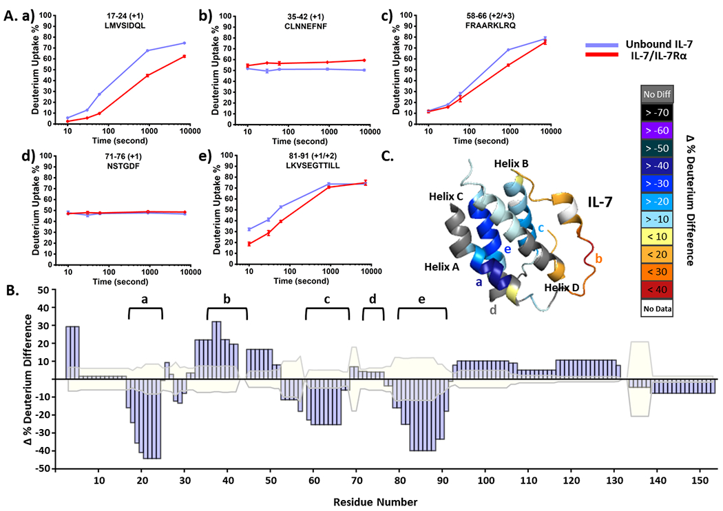
Figure 1.

(A) Representative HDX kinetics of unbound IL-7 (purple) and of bound with IL-7Rα (red). (B) Statistical analysis of cumulative HDX difference for each residue. Residues are considered being affected upon binding with IL-7Rα when the difference is greater than three times the propagated error (shaded in faint yellow) of all time points. (C) The cumulative HDX difference of each residue mapped onto the crystal structure of IL-7 (PDB:3DI2).
