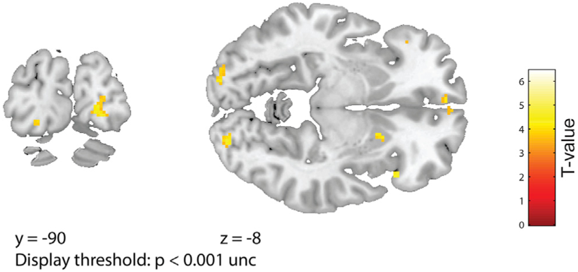
Figure 4. 4-Fold Modulation of fMRI Activity Based on Visual Trajectory in Bilateral V1.

A visual model, based on the up and down vertical movements of the visual bars during the experiment, significantly modulated activity with 4-fold symmetry in bilateral primary visual cortex in left V1 (−20/−88/−14, Z = 3.79; puncorr < 0.0001; small-volume correction [SVC] using an anatomical mask of Brodmann areas 17 and 18: pSVC-corr = 0.003) and in right V1 (22/−90/−8; Z = 3.88; puncorr < 0.0001; pSVC-corr = 0.012). Activations are overlaid on coronal (left) and axial (right) sections of T1 template, at a display threshold of p < 0.001 uncorrected.
