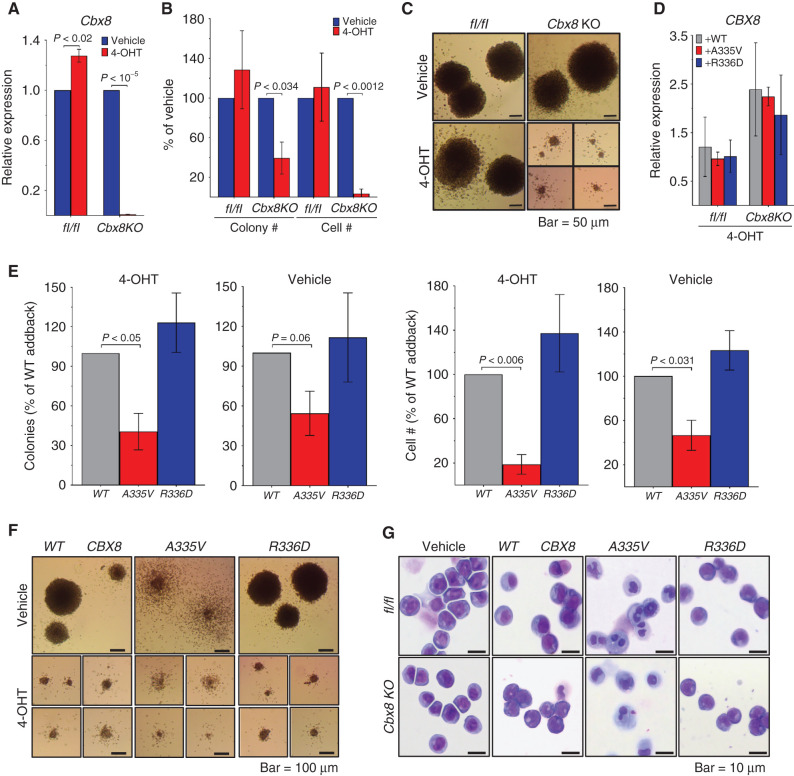
Figure 2.
CBX8 A335V inhibits colony formation, and CBX8 R336D has no effect. MLL-AF9–transformed mouse BM cells from Cbx8fl/fl (flox/flox) and conditional Cbx8 knockout (Cbx8KO) mice after methylcellulose culture 7 days with vehicle or 4-hydroxytamoxifen (4-OHT). For A, B, D, and E, data are from two experiments, each in triplicate, and are represented as mean ± SEM. Statistical significance was determined using a Student two-tailed t test with a confidence interval of 95%. A, Quantification of endogenous Cbx8 RNA expression, normalized to Polr2a by qRT-PCR. B, Quantification of colony and cell number relative to vehicle control. C, Bright-field images of colonies treated with vehicle (top) or 4-OHT (bottom). D, Quantification of exogenous CBX8 WT (gray), A335V (red), and R336D (blue) RNA expression, normalized to Polr2a by qRT-PCR. E, Number of colonies (panels 1 and 2) and cells (panels 3 and 4) produced by conditional Cbx8 knockout mouse BM cells treated with 4-OHT (panels 1 and 3) or vehicle (panels 2 and 4) and expressing exogenous CBX8 WT (gray), A335V (red), or R336D (blue). F, Bright-field images of conditional Cbx8 knockout mouse BM cells expressing CBX8 WT (left), A335V (middle), or R336D (right) and treated with vehicle (top) or 4-OHT (bottom). G, Images of Wright–Giemsa stained flox/flox (top) and conditional Cbx8 knockout (bottom) mouse BM cells treated with vehicle (panel 1) or 4-OHT and expressing exogenous CBX8 WT (panel 2), A335V (panel 3), or R336D (panel 4).

