Fig. 1.

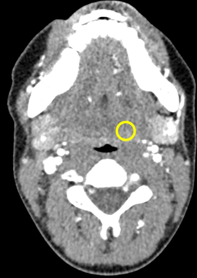
Neck computed tomography image of a patient that was used as a template for the phantom shape. The region of interest in the left parapharyngeal space indicates the position for insertion of low‐contrast signals.

Neck computed tomography image of a patient that was used as a template for the phantom shape. The region of interest in the left parapharyngeal space indicates the position for insertion of low‐contrast signals.