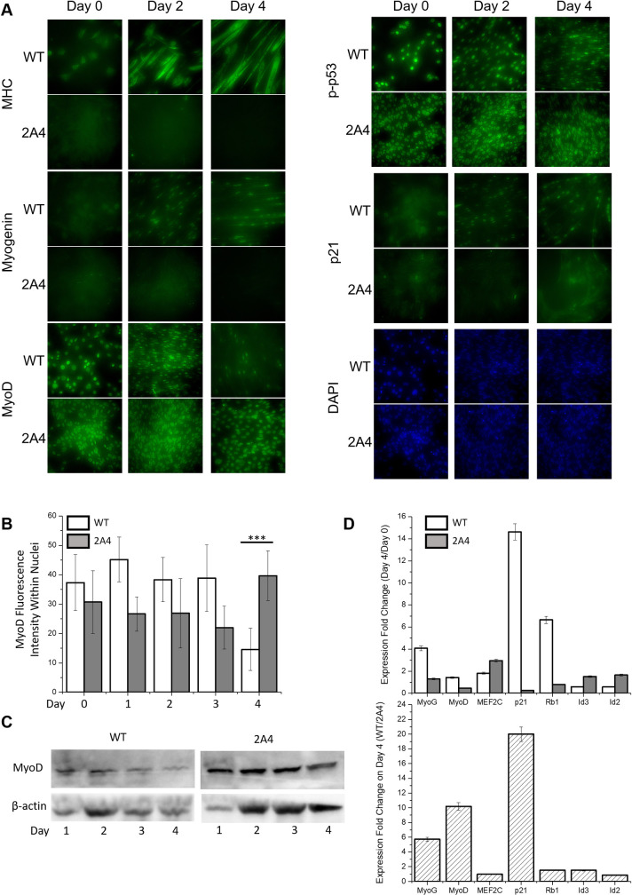
Fig 3. Expression and localization of myogenic regulatory factors in wild type and CRISPR-edited C2C12 myoblasts during myogenesis.
(A) Immunofluorescence to determine the localization of MyoD, p-p53, p21, myogenin, and MHC in differentiating myoblasts. (B) Average fluorescence intensity of MyoD within nuclei of WT and 2A4 cells on days 0 to 4 of differentiation (average of 10 cells per field, n = 2). Data are represented as the mean ± SD; ***P < 0.001. (C) Western blot of WT and 2A4 cell lysates on days 1 to 4 of differentiation (n = 1) to measure MyoD protein level. (D) Fold-change in gene expression measured by RT-qPCR for the indicated genes, calculated as the ratio of gene expression on day 4 to day 1 for WT and 2A4 cells (top panel) and as the ratio of WT to 2A4 cells on day 4 of differentiation (bottom panel) (n = 3). Data are represented as the mean ± SD.

