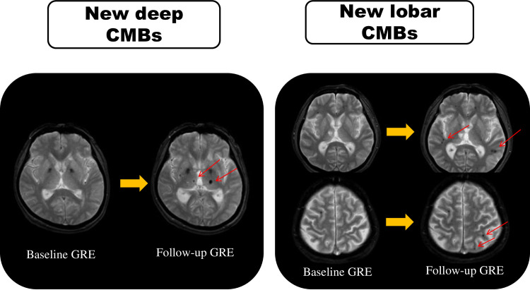
Fig 2. An illustration of MRI in the two patients who showed different types of new CMB locations.
For each patient, the baseline GRE is on the left, and the follow-up GRE is on the right. Arrows indicate new CMBs on the follow-up scan. CMBs: cerebral microbleeds. GRE: T2*-weighted gradient-recalled echo imaging.

