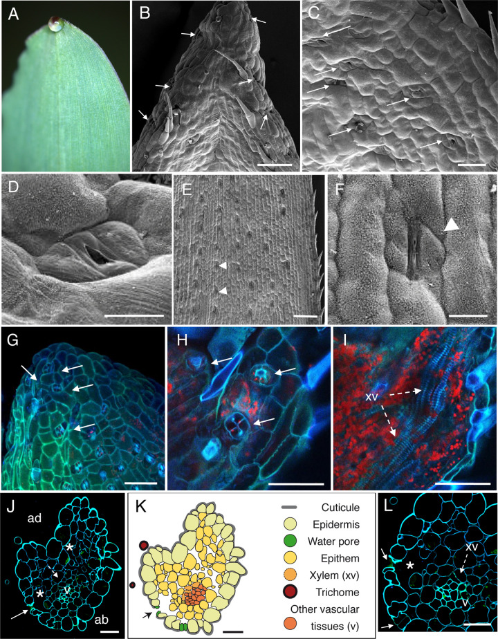
Fig 1. Anatomic description of maize apical hydathodes by confocal and Scanning Electron Microscopy (SEM).
(A) A guttation droplet at the leaf tip. (B-D) The adaxial face of leaf tip was imaged by SEM. Water pores are observed in the gutter. (E-F) Observations of stomata were performed on distal parts of leaves relative to panels B-D. Panel F is a closeup image of panel E. (G-I) Confocal images of fresh adaxial face of the leaf tip. (G) The image is a maximal projection of 50 confocal planes in z dimension (1-μm steps). (H-I) Observations in z axis of the same sample at the epidermal level (H) and below the epidermal layer (I). Each overlay image corresponds to the maximal projection of 25–30 confocal planes acquired in z dimension. (J, L) Transversal sections (1-μm thickness) of fixed tissue at 80–100 μm from the tip were observed by confocal microscopy. White arrows, arrowheads, dashed arrows and asterisks indicate water pores, stomata, xylem vessels (xv) and large chambers and intercellular spaces, respectively. v, small veins; ad, adaxial face; ab, abaxial face. (K) Schematic drawing of the hydathode cross section observed in J. Scale bars: B, E, G-I: 100 μm; F: 20 μm; C: 40 μm; D, J-L: 30μm.

