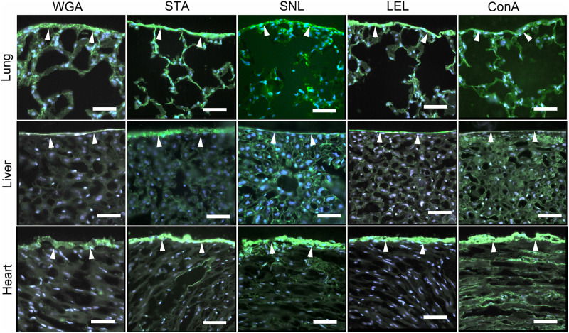
Fig 1. Fluorescent lectin staining of the mesopolysaccharides of the lung, heart and liver.
Frozen tissue sections were warmed to 27°C before fixation in 2% paraformaldehyde. The lectin fluorescence detection was performed as described in Materials and Methods and images were obtained with fluorescent microscopy. The lectin staining was demonstrated with fluorescently labeled Concavalia ensiformis (ConA) [18], Lycopersicon esculentum lectin (LEL) [19], Sambucus nigra (SNL) [20], Solanum tuberosum (STA) [21] and wheat germ agglutinin (WGA) [22].

