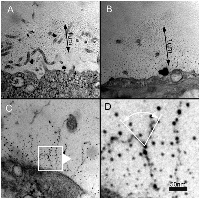
Fig 2. Chemical fixation, ruthenium red staining and transmission electron microscopy of the murine visceral pleura.

A,B) The ruthenium-stained carbohydrate layer appears to extend approximately 1um from the pleural mesothelial surface. C,D) Detailed analysis of the images suggested that the MPS is composed of beaded strands with a branching structure.
