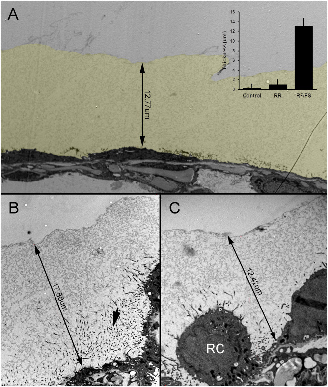
Fig 3. HPF preservation of the MPS expressed on the murine visceral pleura.
A) The MPS was several-fold thicker than the underlying mesothelium and significantly thicker than the glycocalyx detected by conventional ruthenium red (RR) staining (control) (inset p<.0001). The MPS is pseudocolored yellow for presentation purposes. B-C) HPF fixation of the visceral pleura demonstrated MPS thickness ranging from 8um to 18um. Microvilli, seen in cross-section, extended into the MPS from the associated mesothelium (B, arrow). Notably, the MPS was present even in the setting of activated or reactive (RC) underlying mesothelial cells [25].

