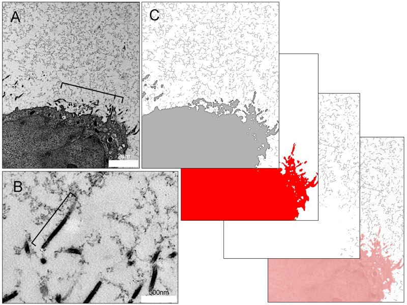
Fig 4. Mesothelial microvilli anchoring the MPS of the visceral pleura.
A) HPF TEM of mesothelial cell microvilli (bracket) and surrounding MPS in tissues preserved by HPF. B) Magnification of the microvilli demonstrates traces of the MPS linked to the microvilli (bracket). Cross-section of the pleura imaged with TEM. The microvilli extend 1.1 ± 0.35 um from the mesothelial surface. C) Image analysis using a skeletonization algorithm illustrates the branched-chain structure and connectivity of the carbohydrate biopolymer with an average branch number of 2.77 branches, a mean branch length of 134.86 ± 81.70 nm and a Euclidean distance of 115.65 ± 65.46 nm between skeletonized branches.

