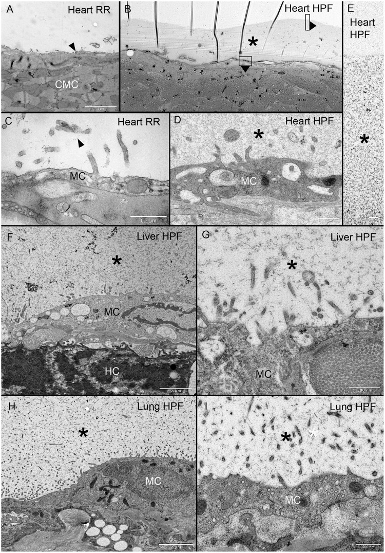
Fig 5. MPS expressed on heart, liver and lung organ surface.
Ruthenium red staining resulted in scant contrast detected on the heart surface (A, C arrows). In a light microscopic overview of a 299 nm thick section stained with toluidine blue, the murine heart surface demonstrated a much thicker surface layer (B, asterisk). TEM images of similar areas of the MPS is shown (D, E asterisk). Surface features of the MPS were also similar on the liver (F, G asterisk) and lung (H, I asterisk). Note the similar branched-chain structure in all three organs. RR = ruthenium red; HPF = high-pressure fixation; MC = mesothelial cell; CMC = cardiac myocyte; HC = hepatocyte. Asterisk identifies the MPS in all tissues.

