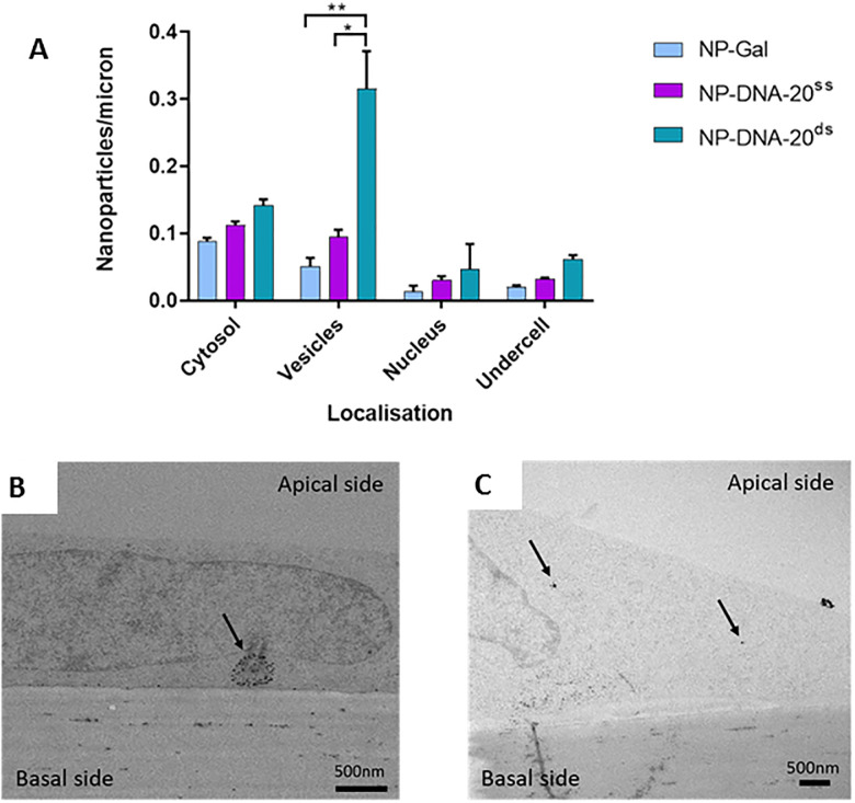
Fig 2. Endocytosis of gold nanocarriers with ssDNA or dsDNA by brain endothelium.
(A) Nanoparticles in cytosol, vesicles and the nucleus of hCMEC/D3 cells. NPs with 20nt ssDNA (NP-DNA-20ss) were compared with dsDNA(NP-DNA-20ds) and the base nanoparticles (NP-Gal). Three experiments were performed, each individual experiment with three technical repeats. Tukey's multiple comparisons test showed a significant difference for NP-DNA-20ds compared to NP-Gal (** P = 0.0034) and NP-DNA-20ss (* P = 0.0133) in vesicles. (B) Transmission electron micrographs showing examples of silver enhanced gold NPs in vesicles (NP-DNA-20ds, A) or the cytosol (NP-Gal, B) of hCMEC/D3 cells after application to the apical surface.

