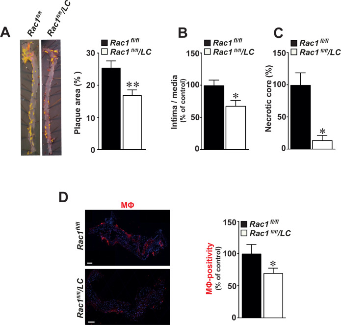
Fig 3. Mice lacking RAC1 in macrophages develop smaller aortic atherosclerotic plaques.
(A) Representative images of Sudan IV-stained atherosclerotic aortic plaques in aortas obtained from Rac1fl/fl/LC mice infected with AdPCSK9 (n = 12) as compared to Rac1fl/fl control mice infected with AdPCSK9 (n = 12) (left panels). Quantification of atherosclerotic plaque size by image analysis in Rac1fl/fl and Rac1fl/fl/LC aortas infected with AdPCSK9 (right graphs). (B) Intima/media ratios in Rac1fl/fl and Rac1fl/fl/LC aortic arches from mice infected with AdPCSK9. (C) Percentage of intimal necrotic core areas in Rac1fl/fl and Rac1fl/fl/LC aortic arches. (D) Immunofluorescent detection of macrophages (MΦ) using anti-CD68 antibodies in Rac1fl/fl and Rac1fl/fl/LC aortic arches from mice infected with AdPCSK9 (left panels). Number of MΦ in Rac1fl/fl and Rac1fl/fl/LC aortic arches (right graphs). Scale bars represent 100 μm. Mean ± SEM values. Student’s t-test was used. *p≤0.05.

