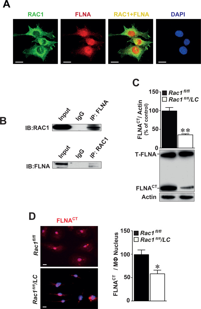
Fig 6. RAC1 interacts with FLNA in the cytoplasm and deletion of RAC1 reduces the production of cleaved C-terminal fragment of filamin A (FLNACT).
(A) Expression of RAC1 (green) and FLNA (red) is co-localized mainly in the cytoplasm of BMMs (yellow) as detected by immunofluorescence staining. Scale bar represents 10 μm. (B) Co-immunoprecipitation identifying FLNACT as an interacting partner of RAC1. Total proteins obtained from BMMs immunoprecipitated with FLNACT and then immunoblotted with RAC1 antibodies (upper) or vice versa (lower). IgG served as negative controls. Full-length of FLNA and FLNACT were indicated and actin served as internal loading control. (C) Immunoblotting of FLNA in Rac1fl/fl and Rac1fl/fl/LC BMMs. (D) Immunofluorescence staining of FLNACT in Rac1fl/fl and Rac1fl/fl/LC BMMs. Quantification of nuclear FLNACT expression in Rac1fl/fl or Rac1fl/fl/LC BMMs. Scale bar represents 20 μm. Mean ± SEM values of at least quadruplicated experiments. Student’s t-test was used. *p<0.05, **p<0.01.

