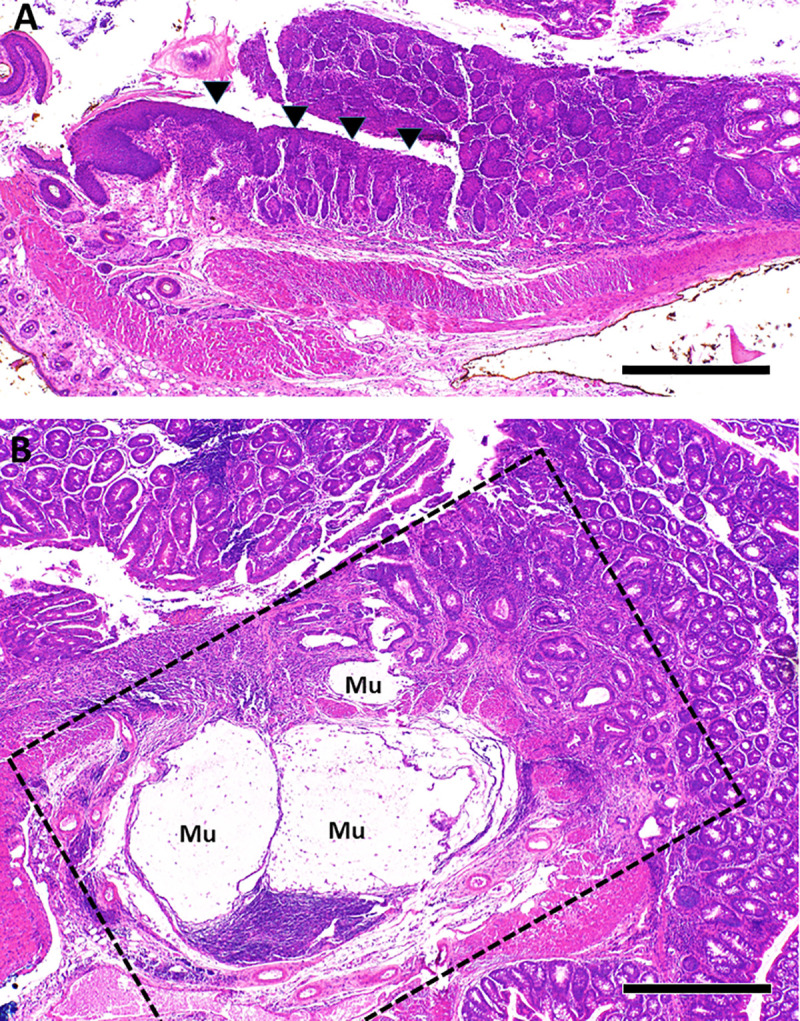
Fig 2. Squamous metaplasia and neoplasia in TIA mice.

A. Squamous metaplasia (arrowheads) was common in the rectum of TIA mice, shown here in a 9 week old. B. The dotted lines highlight a focus of invasive mucinous adenocarcinoma identified in the proximal colon of a 15 week old TIA mouse with severe pan-colitis (histologic score = 55). “Mu” indicates mucin lakes associated with invasive carcinoma. Scale bar in A and B represents 500 μm.
