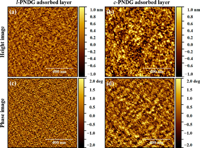
Figure 9.

Representative AFM height images of the (a) l-PNDG and (b) c-PNDG adsorbed layers. The corresponding phase images are shown in (c) and (d), respectively. The scales of height and phase images are ±1 nm and ±2°, respectively. The scan size of all images is 1 μm × 1 μm.
