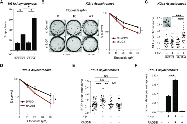
Figure 7.
Homologous recombination prevents chromosomal instability induced by TOP2. (A) Apoptosis analysis by FACS of KG1a shRNA LIG4 and control cells. Asynchronous cells were treated with 40 μM etoposide for 3h and incubated in drug-free medium for 48 h after treatment. Mean (± s.e.m.) of the percentage of Annexin-V positive cells of three independent experiments. Statistical significance was determined by t-test (*P< 0.05). (B) Clonogenic survival of KG1a shRNA LIG4 and control cells treated with the indicated concentration of etoposide for 3 h. After treatment, cells were washed and cultured in methylcellulose-containing medium for 14 days. Left, representative images of cultures. Right, quantification (mean ± s.e.m.) of three independent experiments. Statistical significance was determined by two-way ANOVA (*P< 0.05). (C) Sister chromatid exchanges of KG1a shRNA LIG4 and control cells treated with 10 μM etoposide for 30 min. After that, cells were washed and cultured in drug-free medium for 8 h, previous to metaphase spread preparation. See insets for representative image of SCEs. Mean (± s.e.m.) of SCE events per chromosome per metaphase from two independent experiments. Statistical significance was determined by t-test (***P< 0.001). (D) Clonogenic survival of RPE-1 cells with the indicated concentration of etoposide for 3 h. Where indicated cells were pre-incubated with RAD51 inhibitor (10μM) for 30 minutes prior to, during and 20 h after etoposide treatment. After treatment, cells were cultured in serum containing drug-free medium for 8 days. Data are the mean (± s.e.m.) of three independent experiments. Statistical significance was determined by two-way ANOVA (*P< 0.05). (E) Sister chromatid exchange of RPE-1 treated with 5 μM etoposide for 30 min. Where indicated cells were pre-incubated with RAD51 inhibitor (10 μM) for 30 min prior to, during and after etoposide treatment. Cells were cultured in drug-free medium for 6 h previous to metaphase spread preparation. Mean (± s.e.m.) of SCE events per chromosome per metaphase from two independent experiments. Statistical significance was determined by T-test (**P < 0.01, ***P< 0.001, NS, not significant). (F) Translocation frequencies (translocations per metaphase) in chromosome 8 and 11 were quantified in asynchronous RPE-1 cells in metaphase spreads prepared 24 h after etoposide treatment (1 h, 25 μM). Where indicated, cells were pre-treated with RAD51 inhibitor (10 μM) for 30 min prior to, during, and 6 h after etoposide treatment. Data are the mean (± s.e.m.) from three independent experiments. Statistical significance was determined by t-test (***P< 0.001).

