Figure 3.

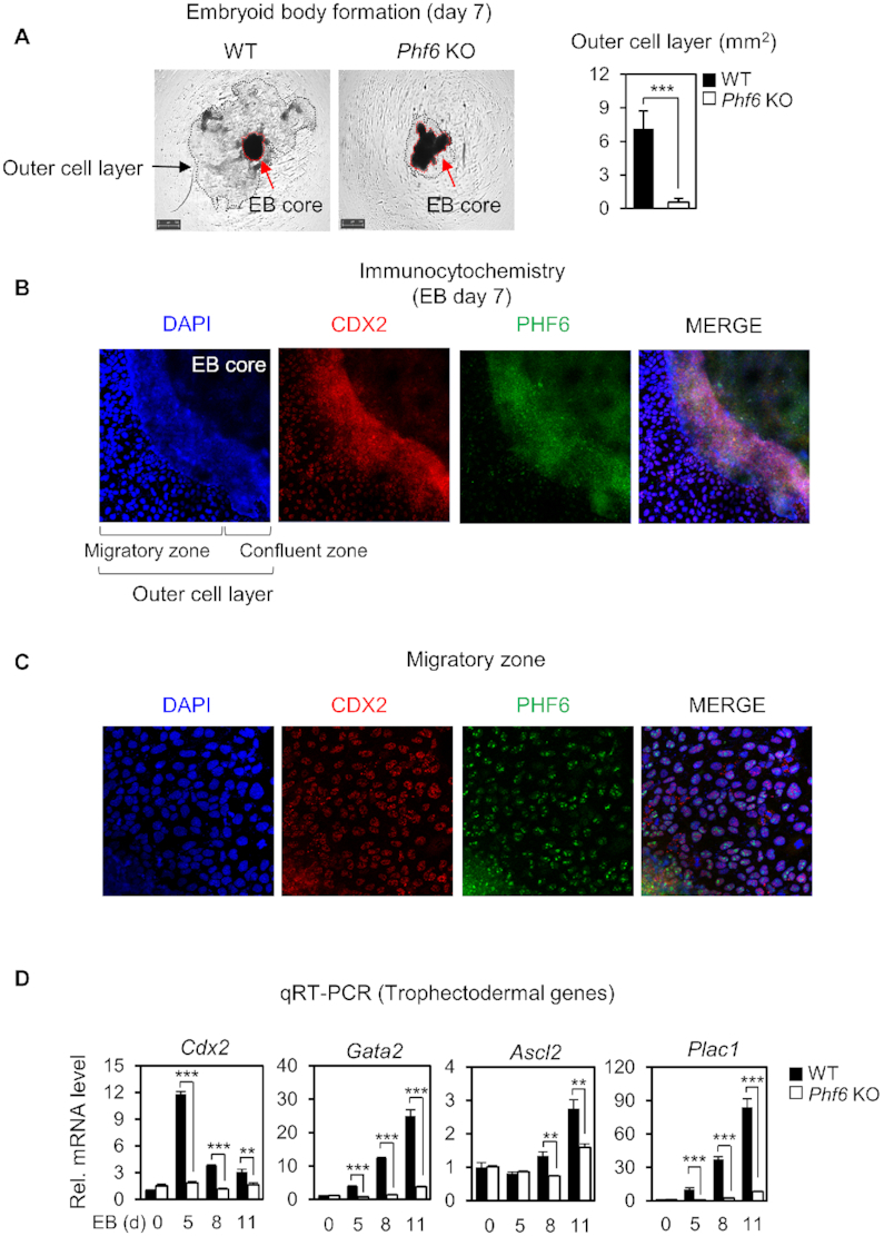
PHF6 activates trophectodermal genes for trophectoderm lineage determination. (A) Morphological features of EBs from WT and Phf6 KO ESCs on Day 7 (left). Each EBs were grown and observed separately. Magnification ×10. The average areas (mm2) of the outer cell layer were compared between WT (n = 11) and Phf6 KO (n = 9) EBs (right). Statistical significance was calculated by ANOVA test (*P < 0.05, **P < 0.01, ***P < 0.001). (B, C) Immunostaining of CDX2 and PHF6 in the outer cell layer in WT EBs. Magnification ×100 (B) and ×200 (C). (D) qRT-PCR analysis of the trophectodermal genes during EB differentiation in WT and Phf6 KO ESCs. WT and Phf6 KO EBs were maintained without LIF and harvested at the indicated days. mRNA levels of each gene were determined as relative values for Gapdh or β-actin and relatively compared based on WT day 0. Statistical significance was calculated by ANOVA test (*P < 0.05, **P < 0.01, ***P < 0.001).
