Figure 4.
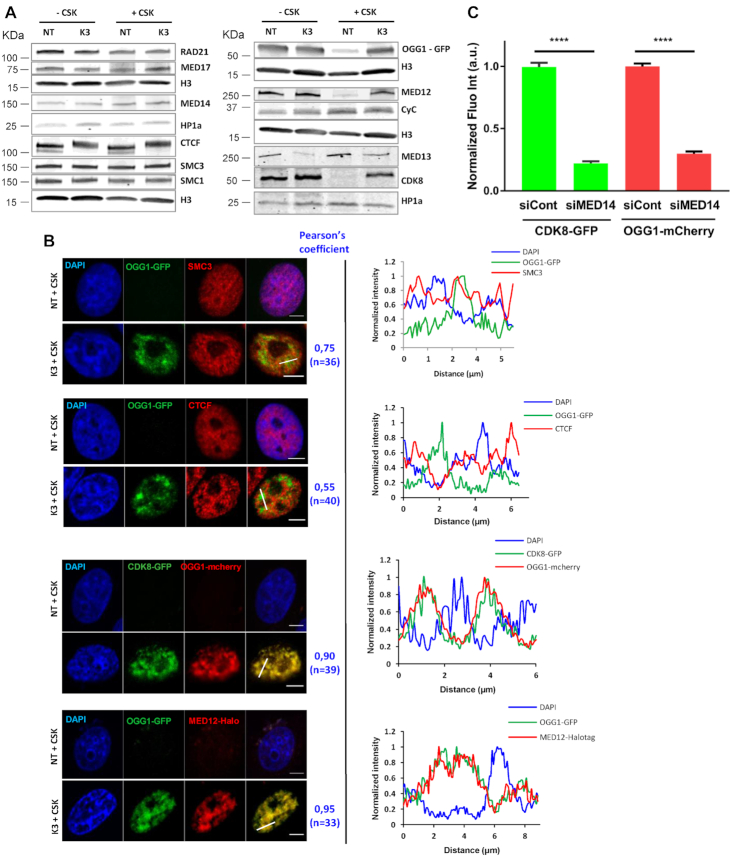
Co-recruitment of mediator CKM subunits and OGG1 to the same nuclear regions. (A) Subcellular fractionation of non-treated (NT) and KBrO3 (K3)-treated OGG1–GFP cells. Whole cell extract (–CSK) or the insoluble fractions (+CSK) were analysed by western blot using the indicated antibodies. HP1α or H3 were used as loading controls. (B) Distribution patterns of OGG1–GFP, SMC3, CTCF, CDK8-GFP and MED12-Halotag in non- (NT) and KBrO3- (K3) treated cells. Prior to fixation, soluble proteins were removed with CSK. Scale bar: 5 μm. Pearson correlation coefficient was calculated for the indicated number of cells from at least two independent experiments. Plot profiles along the lines in the merged image are shown. (C) Quantification of OGG1-mCherry and CDK8-GFP fluorescence intensities after KBrO3 treatment and CSK washing in control cells or cells depleted for MED14. More than 4000 cells were analysed from three independent experiments. Error bars indicate SEM. Statistical analysis involved a Kruskal–Wallis test. (****) P < 0.0001.

