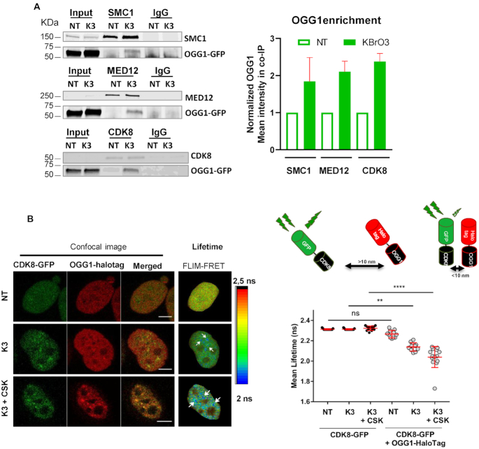
Figure 6.
Oxidative stress induces the association of OGG1 with mediator and cohesins. (A) Immunoprecipitation of SMC1, MED12 and CDK8 was performed in non- (NT) or KBr03- (K3) treated HeLa cells expressing OGG1–GFP after benzonase treatment of the cell extracts. The presence of OGG1, SMC1, CDK8 and MED12 in different fractions (Input, IP using an antibody against the target protein or a control IgG) was evaluated by western blot using specific antibodies. Enrichment of OGG1 in the co-immunoprecipitates was quantified from 3 (MED12) or 2 (SMC1 and CDK8) independent experiments. Mean intensity measured was normalized to the amount detected in extracts from NT cells set to 1. Error bars represent SEM. (B) Intracellular distribution of OGG1-Halotag and CDK8-GFP fusion proteins in non- (NT) and KBrO3- (K3) treated HeLa cells. Prior to fixation, soluble proteins were removed with CSK as indicated. Scale bar: 5 μm. The spatial distribution of the mean fluorescence lifetime of the GFP donor is displayed using a continuous pseudocolor scale ranging from 2 to 2.5 ns. The graph corresponds to the quantification of donor's fluorescence lifetime in NT and K3 cells expressing the donor alone or the donor and the acceptor. Values obtained for 10 cells from a representative experiment are shown. The results were confirmed in three independent experiments and using mCherry as the acceptor instead of HaloTag (see Supplementary Figure S6). Statistical analysis involved a Kruskal–Wallis test. (****) P < 0.0001.

