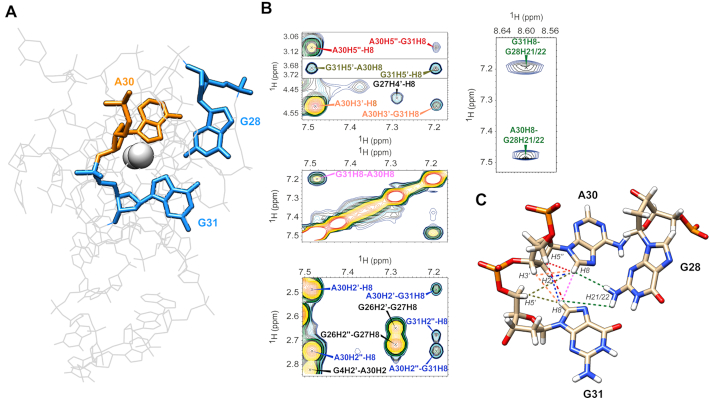
Figure 6.
Representation of the triad within the KRAS 32R G9T calculated structure in (A) with guanines 28 and 31 in blue and adenine 30 in orange. Ten cross peaks proving the existence of the triad at 37°C found in KRAS 32R G9T {1H–1H} NOESY spectrum are show in (B) involving aromatic and sugar protons from the three bases implicated in the triad and the nearby residues. The corresponding restraints are shown in the calculated triad in (C) with the corresponding colors.

