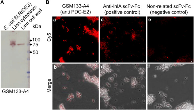
Figure 3.
Test of GSM133-A4 in immunoblot of Listeria protein fractions and immunofluorescence microscopy. (A) Immunoblot of GSM133-A4 of protein fractions from the cytoplasm and cell wall of L. innocua DSM20649 (Linn), as well as E. coli BLR (DE3) extract used as a negative control. The blot shows that GSM133-A4 antibody binds to a target contained both in the cytoplasm and cell wall fractions of Listeria. The used protein ladder was Precision Plus Protein All Blue (Bio-Rad). (B) Fluorescence microscopy on alive L. monocytogenes 4b DSM 15675. The signal of GSM133-A4 (c and f) showed to be higher than both the negative control with a non-related antibody (a and d) and the anti-InlA positive control (b and e). The complete immunoblot image can be found as Supplementary Fig S9.

