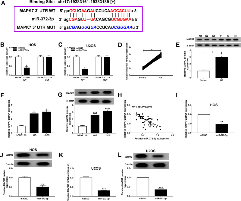
Figure 7.
MiR-372-3p directly interacted with MAPK7 in BC cells. The HOS and U2OS cells were co-transfected with MAPK7 3ʹUTR-WT+miR-NC, MUC19 3ʹUTR-WT+miR-372-3p, MAPK7 3ʹUTR-MUT+miR-NC, 3ʹUTR-MUT+miR-372-3p. (A) Starbase v3.0 predicted the putative site between miR-372-3p and MAPK7. (B and C) The luciferase activity was measured in HOS and U2OS cells. (D-E) The expression of MAPK7 was measured by qRT-PCR and Western blot in OS tissues. (F and G) The expression of MAPK7 was measured by qRT-PCR and Western blot in hFOB1.19, HOS and U2OS cells. (H) The expression of miR-372-3p was negatively correlated with MAPK7 (R=−0.661). (I and J) The level of MAPK7 was determined using the qRT-PCR and Western blot analysis in HOS cells transfected with pcDNA3.0-miR-372-3p. (K and L) MAPK7 level was determined using the qRT-PCR and Western blot analysis in U2OS cells transfected with pcDNA3.0-miR-372-3p.*P<0.05, **P<0.01, ***P<0.001, **** P<0.0001.

